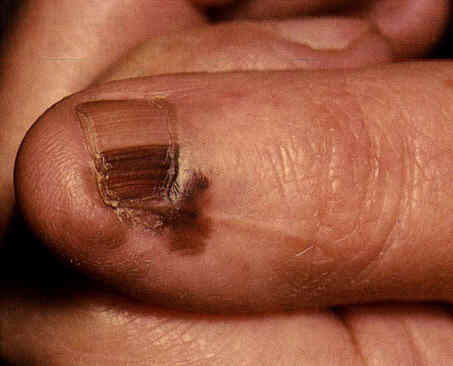
Nail melanoma is a rare type of melanoma that develops in the fingernails or toenails. This type of cancer accounts for about 2% of melanomas in Caucasians and about 30% in African and Asian populations. It affects both men and women equally often, and is more common in people over the age of 50. Often, nail melanoma affects the first finger of the hand more than the others.

Originating from melanocytes, the cells responsible for producing the pigment melanin, nail melanoma appears as a brownish or blackish-brown band under the nail plate. As the disease progresses, the band may become larger and mottled in color, bleeding may appear, and the nail plate may be raised. In some situations, the tumor may also extend to the cuticle, a phenomenon known as Hutchinson’s sign. This sign distinguishes nail melanoma from other nail conditions, such as melanonychia striata, subungual hemorrhage, and nevi, although Hutchinson’s sign is not specific only to melanoma.
In particular, amelanotic melanoma, which is a form of melanoma that lacks pigment, can be particularly difficult to diagnose. In this case, a careful dermatoscopic examination may be necessary to detect areas of peripheral atypical pigmentation or nail plate changes.
In terms of treatment, nail melanoma often requires an incisional biopsy, which involves removing the nail matrix, or an excisional biopsy. In more advanced cases, it may be necessary to remove the entire finger and potentially regional lymph nodes. It is also very important to watch out for pyogenic granuloma of the fingers due to ingrown toenail, as it could actually be amelanotic melanoma.
As with all types of melanoma, early diagnosis and prompt treatment are key to improving your chances of survival. Therefore, it is important to see a doctor if you notice any unusual changes in your nails, particularly if a nail band does not progressively disappear, becomes enlarged, and there is no previous trauma.